Complete Anatomy
Complete Anatomy (CA) is a powerful, highly detailed anatomy visualization platform available via mobile, tablet, and laptop. Used by Stanford Medical Students as an anatomical learning aid, CA provides 3D models of human anatomy available as female and male, allowing for contrasting different anatomies between sexes. Like a “virtual” dissection lab, layers can be removed and anatomical structures — musculoskeletal, neurovascular, digestive, urogenital, and respiratory systems – can be explored.
Key Features Include:
Atlas
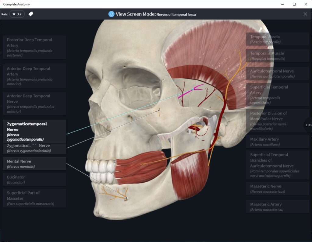
The CA Atlas includes over 700 pre-set anatomical dissections, e.g., Nerves of the Temporal Fossa (Fig 1)
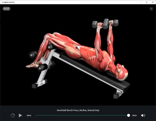
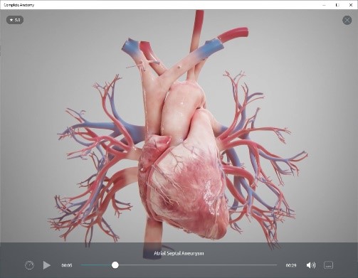
Videos
Animated videos of a wide range of topics related to fitness (e.g., weight lifting, Fig 2), ophthalmology, orthopedics, and cardiology. (Fig 3)
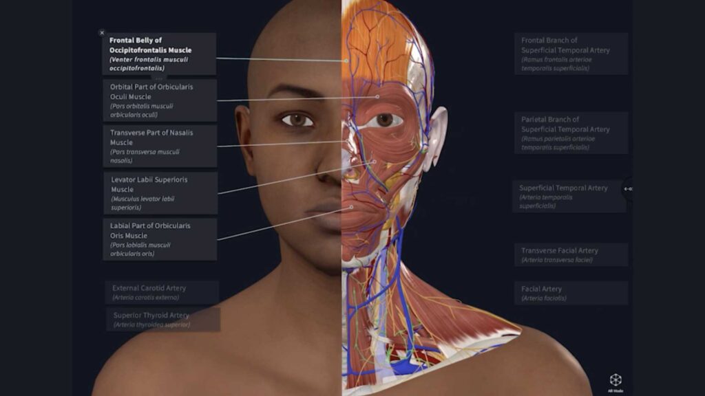
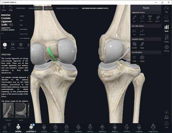
3D Models
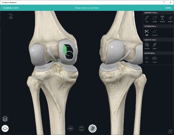
Highly detailed anatomical models that can be “virtually” dissected/rebuilt by removing/adding “layers” (Fig 4) and/or dissecting with “cutting” tools. (Fig 5)
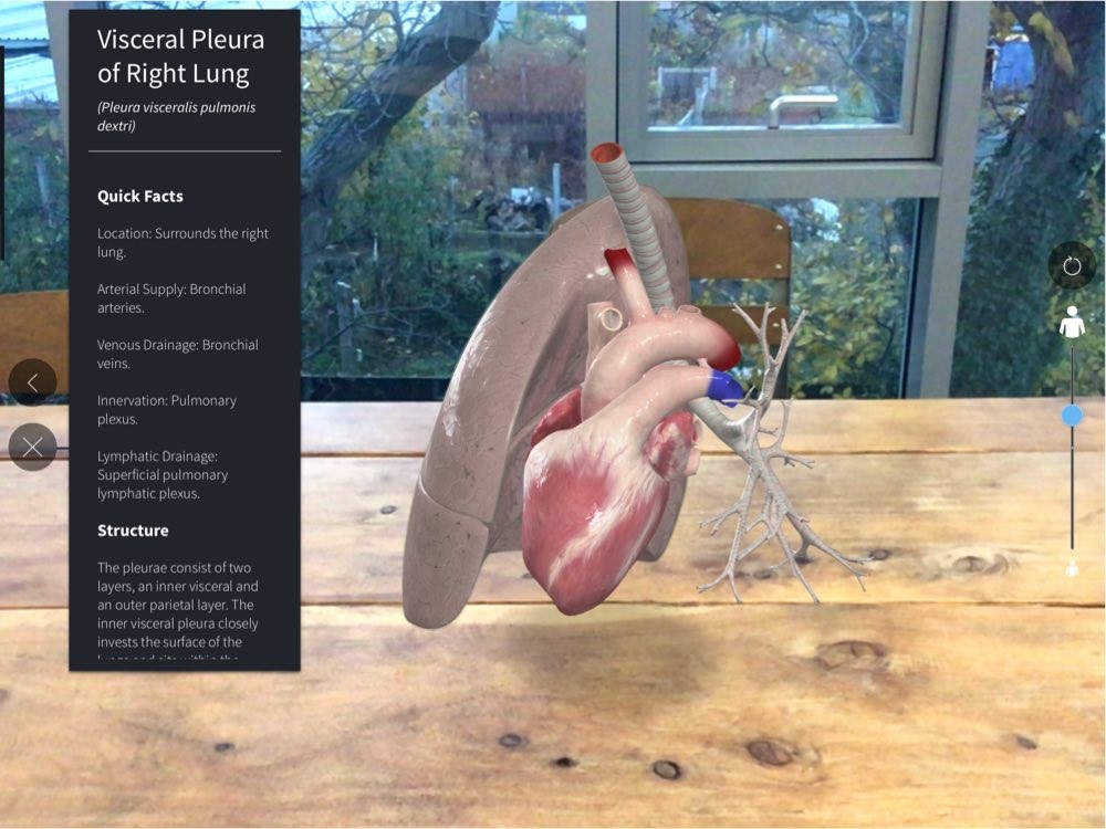
Anatomical objects can be rotated and zoomed in/out, with the images being downloadable. Using the CA mobile app’s Augmented Reality feature, images can be projected via a phone or tablet’s camera onto a flat surface. (Fig 6)
Be sure to explore the amazing richness of this virtual anatomy visualization tool by visiting our Mobile Apps page for instructions on installation and activation.
Need Help?
If you have questions about Complete Anatomy, please explore their support center. You can also explore their Using the Product page for help getting started.