Explore What’s New in Complete Anatomy
Complete Anatomy continues to advance its platform to better support students and researchers in studying and teaching the human body’s complex systems. Find out for yourself and learn why this interactive 3D platform, available through Lane Medical Library, is a favorite resource for medical students, postdocs, and faculty!
NEW: Explore the Brain in 3D
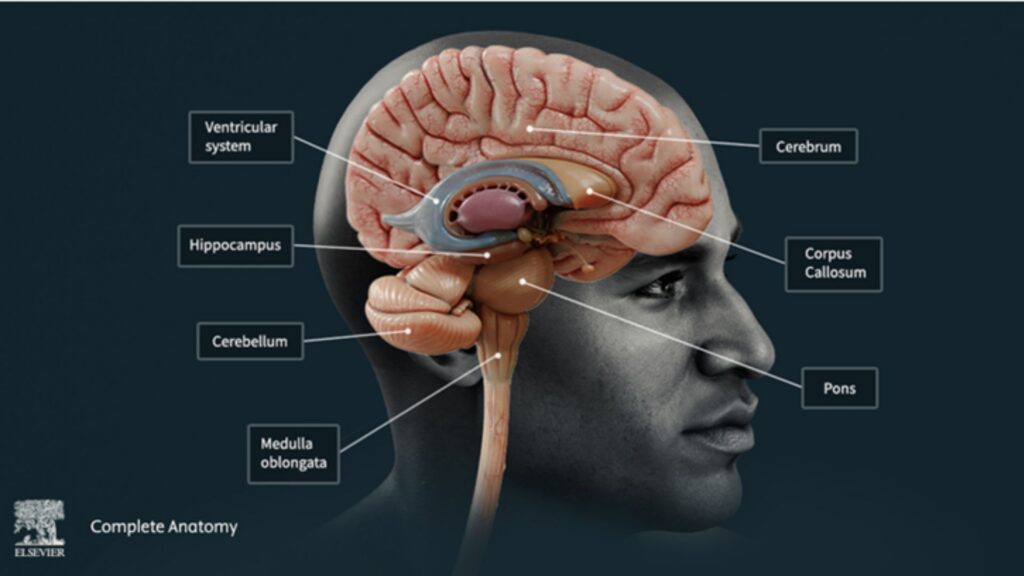
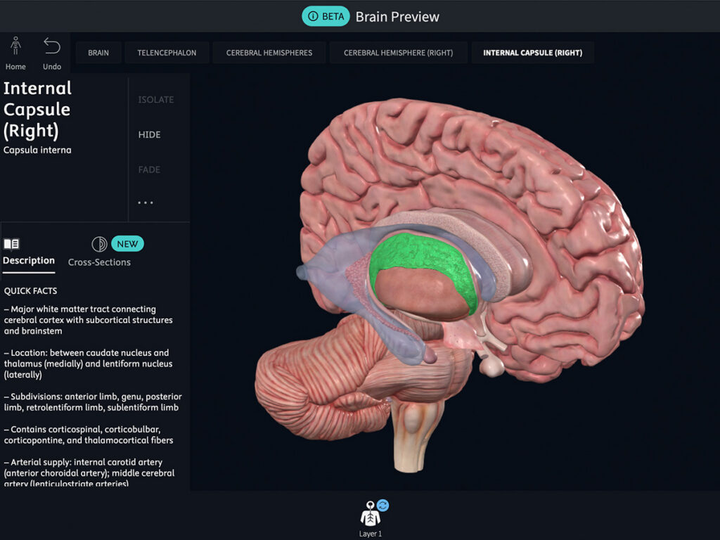
The latest update introduces a new 3D brain model. Explore the gyri with a new brain map and move through coronal, sagittal, and axial cross-sections to better understand neuroanatomy from multiple perspectives, including the structures and regions affected by neurological conditions such as Alzheimer’s disease.
NEW: Expanded Interactive Radiology
Complete Anatomy now offers full-body radiology coverage with hundreds of interactive images aligned with detailed 3D anatomy models. Compare real scans with 3D structures and explore anatomy as it appears in clinical imaging. Get the help you need to recognize anatomical abnormalities, understand how disease can alter normal structures, and connect foundational anatomy with your clinical practice.
Take Complete Anatomy with You on Your Mobile Devices
For mobile access:
- Download Complete Anatomy from the App Store or Google Play.
- Register in the app using your stanford.edu, stanfordhealthcare.org, or stanfordchildrens.org email.
- Go to Settings → My Account and enter the Activation Code.
- Find the Activation Code by searching “Complete Anatomy” on the Lane Library website and opening the Digital Access information.
Whether you’re reviewing anatomy for class or need a quick visual reference for your research, Complete Anatomy puts interactive 3D anatomy and radiology at your fingertips. Contact your liaison librarian with any questions about Complete Anatomy.